-
Categories
-
Pharmaceutical Intermediates
-
Active Pharmaceutical Ingredients
-
Food Additives
- Industrial Coatings
- Agrochemicals
- Dyes and Pigments
- Surfactant
- Flavors and Fragrances
- Chemical Reagents
- Catalyst and Auxiliary
- Natural Products
- Inorganic Chemistry
-
Organic Chemistry
-
Biochemical Engineering
- Analytical Chemistry
-
Cosmetic Ingredient
- Water Treatment Chemical
-
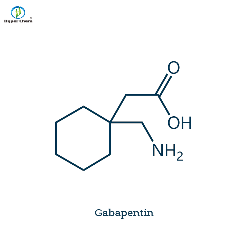
Pharmaceutical Intermediates
Promotion
ECHEMI Mall
Wholesale
Weekly Price
Exhibition
News
-
Trade Service
*Only for medical professionals to read, reference and suggest collections! Common lesions in the brainstem area▌ Brainstem lesions are infarcts, hemorrhages, injuries, tumors, inflammations, metabolic diseases, etc.
▌ Acoustic neuroma, meningioma, epidermoid cyst, craniopharyngioma, etc.
outside the brainstem disease.
▌ Brainstem imaging anatomy The cranium is divided into telencephalon, diencephalon, midbrain, pons, medulla oblongata and cerebellum.
The midbrain, pons, and medulla oblongata are collectively called the brainstem.
The medulla oblongata connects with the spinal cord through the foramen magnum.
▌ Normal CT findings ▌ Normal MRI findings 1 Brainstem infarction ▌ CT findings in hyperacute phase (<24h): negative.
Acute phase (2~6d): low density.
Subacute phase or absorption phase (7~42d): low density.
Chronic phase (﹥42d): CSF-like density.
▌ MRI showed small patches or strips of abnormal signals in the brain parenchyma, with low signal on T1W and high signal on T2W.
The early boundary is blurred, and the late boundary is clear and sharp.
There is no edema around and no mass effect.
▌ Subacute phase infarction 2 brainstem hemorrhage ▌ CT appearance: super acute phase (4-6h): less than 4h, slightly higher density, CT value is about 55Hu, after 4h, the density increases, CT value can be as high as 90Hu.
Acute phase (7-72h): high density, CT value is about 90Hu.
Subacute phase (4d-3w): The density gradually decreases from the periphery.
The diameter of the hematoma is less than 2cm, and it becomes isodensity around 19 days.
The diameter of the hematoma is more than 2cm, and it becomes isodense at about 4-6w.
Chronic phase (3w): cystic or low-density softening lesions, 20% of small hematomas can be absorbed and disappeared.
▌ Acute bleeding ▌ MRI manifestations in hyperacute phase: hematoma is mainly composed of oxygenated hemoglobin, with iso-signal or slightly high signal on T1W; high signal on T2W, and hematoma stratification is not obvious.
Acute stage: Deoxygenated hemoglobin in the hematoma increases and is paramagnetic.
T1W hematoma is still iso-signal, with a low signal edema zone around it; T2W hematoma has a low signal in the center and a band of high signal around it.
Subacute stage: Deoxyhemoglobin evolves into methemoglobin (with strong paramagnetism), and red blood cells begin to dissolve, surrounded by hemosiderin deposit rings, so the hematoma in this stage is complicated with obvious stratification.
Chronic phase: T1W and other signal core layer, high signal nuclear outer layer, equal or low signal peripheral zone; T2W low signal core layer, high signal nuclear outer layer, and high signal peripheral zone, sometimes in the latter two A thin layer of low signal can be seen between them.
Chronic early stage: red blood cells are lysed, deoxyhemoglobin is completely oxidized to methemoglobin, and hematoma has short T1 and long T2 signal characteristics.
Chronic phase: The hematoma is completely absorbed, forming a cyst similar to cerebrospinal fluid, which is manifested as T1W low signal; T2W is high signal.
▌ Pontine cerebral hemorrhage ▌ Pontine hemorrhage chronic hemorrhage period ▌ Pontine hemorrhage 3 The injury is usually related to rotation and the damage is serious.
4 More than 90% of the tumors are gliomas, which are mostly longitudinal and diffuse growth, which can be divided into localized astrocytoma and anaplastic astrocytoma.
The elderly need to exclude metastatic tumors.
▌ Localized astrocytic tumor T1W has low signal, uniform signal and clear boundary.
T2W high signal, uniform signal.
After enhancement, there was no or slight enhancement, and a few moderate enhancement.
No edema or mild edema.
The space-occupying effect is relatively light.
▌ Anaplastic astrocytoma T1W low signal, clear boundary.
Signals such as T2W, a high signal ring can be seen around the stove.
After the enhancement, the ring is strengthened.
Moderate edema is more common around the tumor.
The space-occupying effect is more obvious.
▌ CT manifestations of metastatic tumors are multi-level or high-density on plain scan, and obvious nodular or circular enhancement after enhancement.
MRI showed T1W low signal, T2W high signal, the signal may be uneven, and obvious nodular or circular enhancement after enhancement.
5 MRI manifestations of inflammation: T1W low signal, T2W high signal, enhanced after enhancement.
6 Leukemia-multiple sclerosis▌ MRI manifestations are typically "inclined" or "target-shaped".
T1W frequently sends low signals.
T2W sends high signals frequently.
The number and extent of lesions on DWI showed more.
▌ In multiple sclerosis M 27Y, the lesion is located behind the medulla oblongata, with slightly low signal on T1WI and high signal on T2WI.
The most common tumors in the cerebellopontine angle area outside the brainstem are acoustic neuromas, accounting for about 3/4 of the tumors in this area.
Followed by meningioma, epidermoid cyst, craniopharyngioma.
▌ CT plain scan of acoustic neuroma is slightly low density, MRI examination T1W low signal, T2W high signal.
▌ Meningioma ▌ Epidermoid cyst has low signal on T1W, high signal on T2W, and calcification can be seen around it.
After enhancement, most of the cyst wall and the contents of the cyst are not enhanced.
▌ Medulloblastoma ▌ Craniopharyngioma Source of this article: Yingling Academy Review of this article: Deputy Chief Physician Li Tuming Editor: Mr.
Lu Li Copyright statement This article is reproduced and forwarded to the circle of friends-End-Call for papers.
Welcome to the editor’s mailbox: yxjsjbx@yxj.
For org.
cn, please indicate: [Submission] Hospital + Department + Name The manuscript is in the form of word document, and the remuneration is favorable.
Edit WeChat: chenaFF0911