-
Categories
-
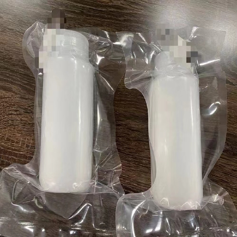
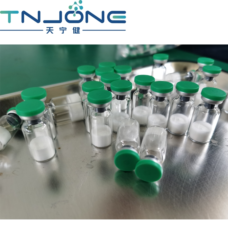
Pharmaceutical Intermediates
-
Active Pharmaceutical Ingredients
-
Food Additives
- Industrial Coatings
- Agrochemicals
- Dyes and Pigments
- Surfactant
- Flavors and Fragrances
- Chemical Reagents
- Catalyst and Auxiliary
- Natural Products
- Inorganic Chemistry
-
Organic Chemistry
-
Biochemical Engineering
- Analytical Chemistry
-
Cosmetic Ingredient
- Water Treatment Chemical
-
Pharmaceutical Intermediates
Promotion
ECHEMI Mall
Wholesale
Weekly Price
Exhibition
News
-
Trade Service
Introduction to Medical Imaging Service Center
As the first WeChat platform in China to open the automatic learning function of medical imaging series, there are hundreds of typical imaging signs, hundreds of imaging case analysis, imaging anatomy, imaging system lectures, foreign excellent lectures, imaging medical affairs and three basic examinations, You can enjoy the knowledge feast with the touch of a finger, such as the work certificate exam and other content, and friends who love learning come to join us
How to join: Enter the Medical Imaging Service Center in the WeChat public account, or click the blue word above to follow it for free
Rheumatoid arthritis (RA) is a chronic inflammatory disease that usually symmetrically affects the facet joints of the hands and feet
We identified 5 patients with RA with pulmonary involvement and performed a retrospective analysis of their chest X-ray and chest CT scans in an attempt to characterize pleural effusion, hydropneumothorax, chylothorax, pulmonary nodular/nodular pleural effusion in such patients Imaging findings of macronodular/necrotizing nodular lesions, pleural plaques, ground-glass opacities, and interstitial lung disease
Case profile
figure 1
(Patient 1): Age 46, male, admitted to hospital with dyspnea and cough
figure 2
(Patient 2): Age 45, male, admitted to hospital with severe chronic cough
image 3
(Patient 3): Age 56, female, admitted to hospital with cough and fever
Figure 4
(Patient 4): 80 years old, male, admitted to hospital for exertional dyspnea
Figure 5
(Patient 5): Age 55, female, admitted to hospital with chronic cough and dyspnea
Among these 5 patients, pulmonary nodules were the most common imaging manifestations of RA, and in the third patient, pulmonary nodules were the first symptom of RA
discuss
In RA patients, the incidence of pulmonary rheumatic nodules is less than 1%, and about 0.
Some pulmonary nodules may have a central necrosis that is hollow and called rheumatic necrotizing nodules
Pleural involvement in patients with RA can manifest as pleural effusion, pleural thickening or pleural plaques, and pneumothorax
According to literature reports, the incidence of RA-related interstitial lung disease fluctuates widely, depending on the method of examination and the population being examined
Pulmonary manifestations of RA also include bronchiolitis, such as follicular bronchiolitis and constrictive bronchiolitis